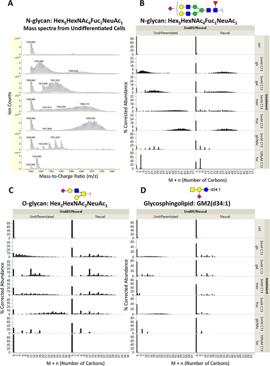
Fig. 2.

(A) Profile mass spectra of the [M + 2H]2+ ion of an N-glycan with composition Hex5HexNAc4Fuc1NeuAc1 from undifferentiated NTERA-2 cells. Because doubly charged species are shown, the mass shift due to 13C labeling is 0.5 Da per labeled carbon. The mass spectra are corrected for the natural isotope distribution of carbon; the corresponding corrected isotopologue data are shown in the first column of (B). Corrected isotopologue data are also shown for the following representative glycans of each type of glycoconjugate: (B) N-glycan, Hex5HexNAc4Fuc1NeuAc1; (C) O-glycan, Hex2HexNAc2NeuAc1; and (D) GSL, GM2(d34:1). Plots are shown for cells grown with regular glucose (ctrl) and [13C-UL] monosaccharides (5 mM glucose, 5 mM galactose, 5 mM mannose, 5 mM fructose and 100 uM fucose).
