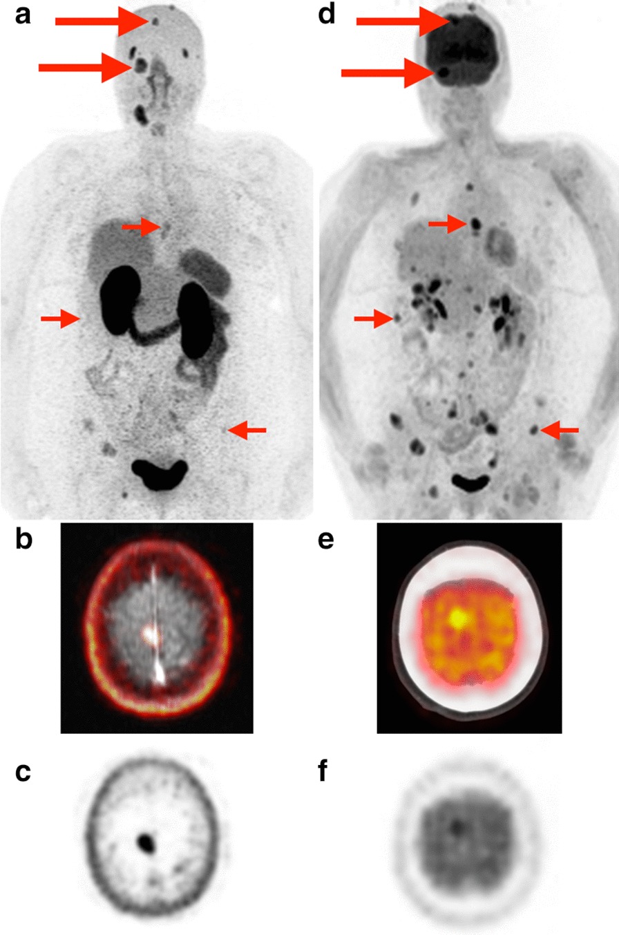
Fig. 5.

Dural and skull base metastases.[68Ga]Ga-PSMA-11 PET MIP (a) and 2-[18F]FDG PET MIP (d) in a patient (Patient 2) with Hurthle cell carcinoma. Dural and skull base metastases (large arrows) are better delineated on PSMA PET due to lack of background brain uptake (example: right falcine dural metastasis better seen on fused axial PSMA PET/MRI (b) and axial PSMA PET (c) than on fused axial FDG PET/CT (e) and axial FDG PET (f). Extensive metastatic disease involving the left adrenal gland, ribs, thoracolumbar spine, and pelvis is better seen on FDG PET in this patient
