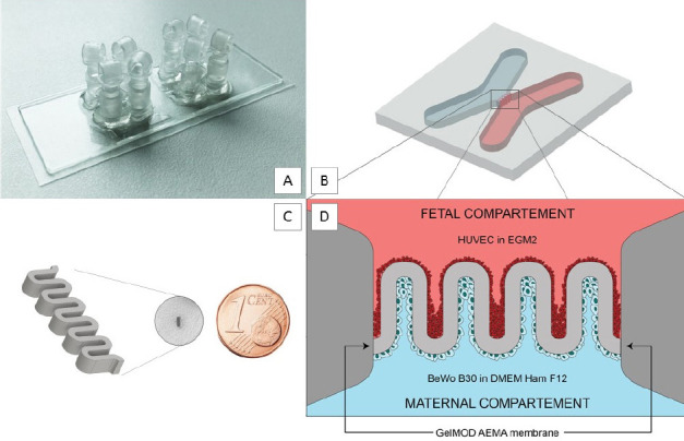
Figure 1.

Placental barrier within a custom-made microfluidic device with two culture chambers.(A) The assembled microfluidic chip with two placental barrier models per slide. (B) The enlarged intersection of the x-shape geometry containing the 3D printed membrane. (C) The CAD-model of the membrane with five consecutive loops mimics the geometry of the placental barrier. To illustrate the size, the structure is shown next to a 1-eurocent coin. (D) After 2PP structuring, the hydrogel membrane separated the chip into two separately perfusable compartments. The fetal and maternal compartment, which were seeded with HUVECs and BeWo B30 cells, respectively. Cells were cultured under constant flow.
