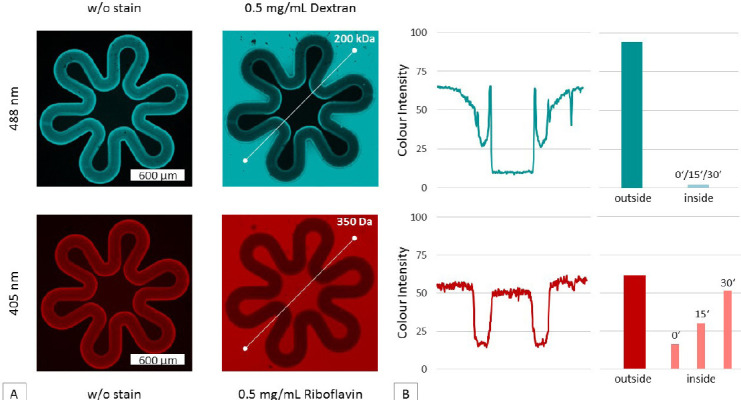
Figure 5.

Test of semi-permeability of a 100 μm thick hydrogel membrane produced with 2PP techniques.(A) In the first column, fluorescence images taken at different wavelengths (488 and 405 nm filter) highlighting the auto-fluorescence of the membranes are shown. In the second column, fluorescence images taken after injection of the stain solutions are shown. The permeability to sugarsized molecules and impermeability to larger molecules is demonstrated using a 0.5 mg/mL riboflavin and 0.5 mg/mL dextran solution. (B) Furthermore, the fluorescence intensity was evaluated along the cross-section of the membrane (white line detail A). Thereby arbitrary units (AU) were measured and plotted using ZEN software. In dextran samples, the inner fluorescence signal is approaching zero. Riboflavin (red) intensity on the other side is equal within and outside the villous structure.
