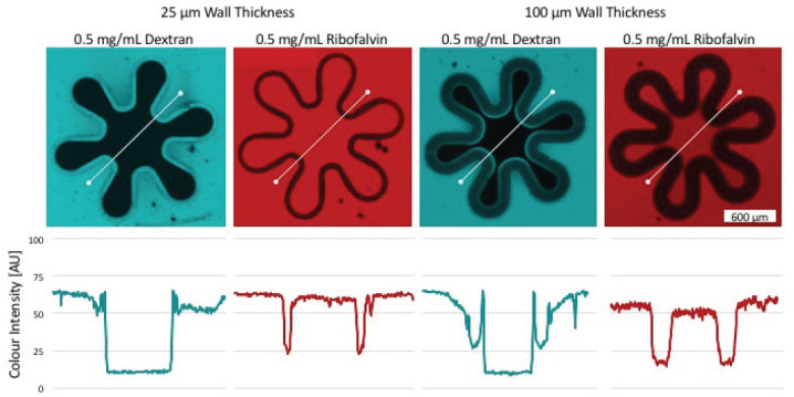
Figure 6.

Color gradient of riboflavin and dextran shows permeability of the membrane with different wall thicknesses.The color intensity was depicted along a linear axis (white). Thereby the color intensity (AU) of pixels on this line was measured and plotted using the ZEN software. Thereby two different wall thicknesses were analyzed. 25 μm on the left side and 100 μm on the right side. The potential to retain high molecular weight substances was demonstrated using dextran (blue) as in both samples the inner fluorescence signal is approaching zero. Riboflavin (red) on the other side is a membrane permeable substance, shown by equal color intensity within and outside the villous structure.
