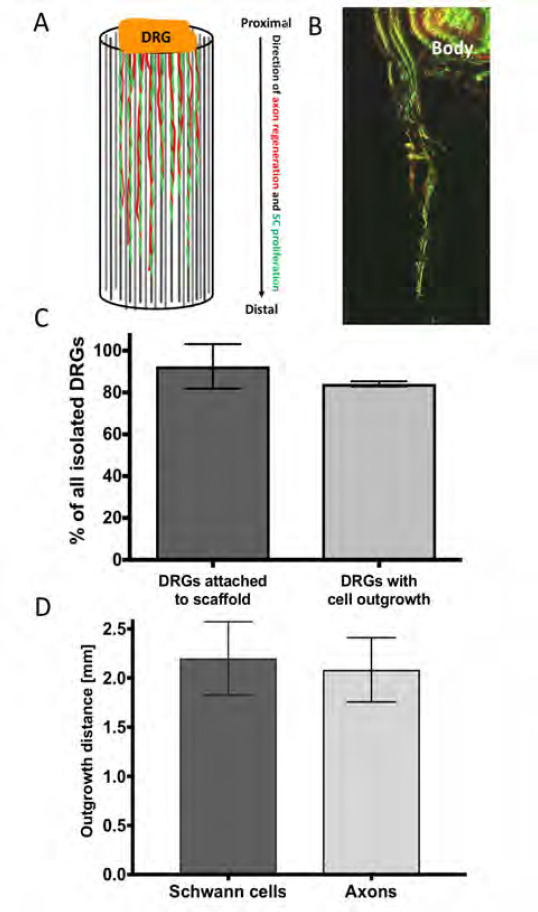
Figure 6.

Ex vivo evaluation of microfibres in whole nerve guides. DRGs were placed on top of the NGC test devices to evaluate the performance of the microfibre scaffold by analysing the outgrowth behaviour of cells along the scaffold. (A) Graphical illustration of the outgrowth of proliferating/ migrating Schwann cells (SC, illustrated in green) and the extension of axons from sensory neuronal cell bodies (illustrated in red) from the DRG body (proximal site) along the microfibres to the tube end (distal site). (B) Confocal microscopy z-projection (depth: ~500 μηι) of the outgrowth of Schwann cells (immunocytochemically-labelled for S100β, green) and axons (immunocytochemically-labelled for βIII-tubulin, red) along PCL microfibres in a 5 mm long PEG NGC. (C) Ex vivo performance of the developed culture setup. From all isolated DRGs 91.6 ± 11.8 % attached to the NGC test device using the proposed setup, from which 85% showed an outgrowth of cells into the test conduit. (D) In the 5 mm long NGC devices, Schwann cells proliferated in average 2.2 ± 0.37 mm into the conduits, where axons grew out 2.1 ± 0.33 mm in 21 days of culture.
