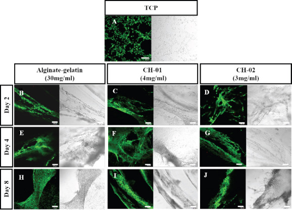
Figure 5.

Live/dead staining of mouse myoblast cells encapsulated in peptide hydrogels, 4 mg/mL CH-01 and 3 mg/mL CH-02, and 30 mg/mL alginate-gelatin (1:1), for different time points. Alginate-Gelatin used as positive control (B, E, H), CH-01 (C, F, I), and CH-02 (D, G, J) at day 2, 4 and 8, respectively. Scale bars 100 μm.
