Figure 2.

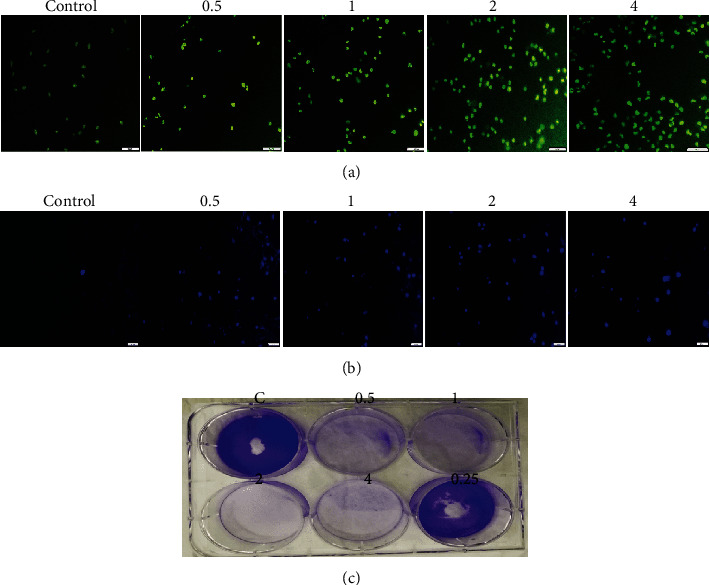
Fluorescent microscopy of HepG2 cells and colony formation assay. (a) AO/EB staining of HepG2 cells after incubation with 0.5, 1, 2, and 4 mg/mL of BLE for 24 h. Live cells stained as uniformly light green in control (untreated) cells, while early apoptotic cells stained as green cells with bright green nuclei that gives the bright green appearance, and necrotic cells/late apoptotic cells stained as light orange. (b) DAPI staining shows the control (untreated) cells as uniformly light blue and the cells after 24 hour of incubation with BLE appeared bright blue with dense nuclei due to chromosomal condensation and nuclear fragmentation as a result of apoptosis. All pictures were taken under the fluorescence microscope (scale bar = 20 μm). (c) The HepG2 cells were grown on a 6-well plate and subsequently incubated with BLE for 24 h, followed by 8-day growth in complete media; it exhibited complete disassociation of colony-forming potential of cancer cells at all high (4, 2, 1, and 0.5 mg/mL) doses and its ability diminishes at 0.25 mg/mL.
