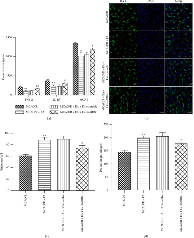
Figure 6.

ABIN1 knockdown impairs the antineuroinflammatory effect of EA. (a) The concentrations of TNF-α, IL-1β, and MCP-1 in the peri-infarct cortex were detected using ELISAs at 24 h after reperfusion (n = 5 rats per group). (b) Microglial morphology was observed by conducting immunofluorescence staining for Iba-1 (green) in the peri-infarct cortex at 24 h after reperfusion (n = 3 rats per group). Scale bar = 50 μm. (c and d) Quantification of microglial endpoints/cell and process length/cell (∗∗P < 0.01 compared to the MCAO/R group; #P < 0.05 and ##P < 0.01 compared to the MCAO/R + EA group).
