Figure 3.

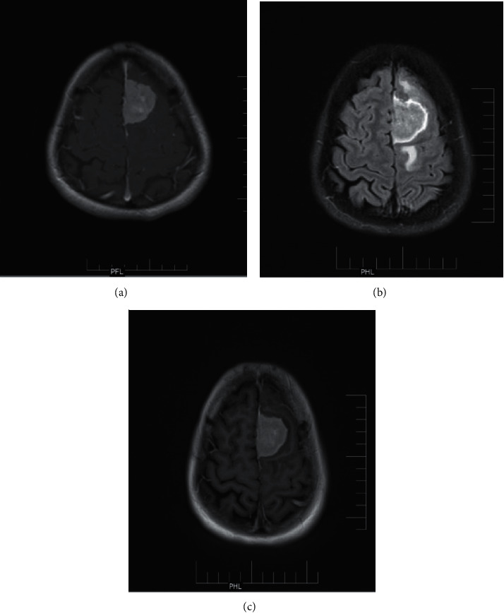
Case of meningioma: (a) postcontrast T1 MTC image; (b) delayed postcontrast T2 FLAIR image; (c) delayed postcontrast T1 FLAIR image.

Case of meningioma: (a) postcontrast T1 MTC image; (b) delayed postcontrast T2 FLAIR image; (c) delayed postcontrast T1 FLAIR image.