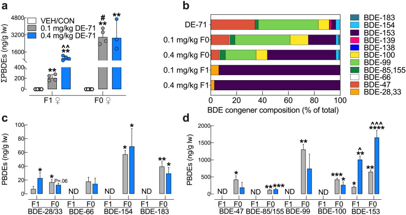
Figure 2.
Hepatic BDE congener analysis of DE-71-exposed mouse dams and their adult female offspring. (a) The ng/g lipid wt sum concentrations of the PBDE congeners detected (bars represent ∑PBDEs; geometric mean ± geometric SD) were comprised of BDE 28/33, BDE-153 for F1 and BDE-28/33, BDE-47, BDE-66, BDE-85/155, BDE-99, BDE-100, BDE-153, BDE-154, BDE-183 for F0. DE-71 exposure produced significant accumulation of PBDEs in liver, being greater in directly exposed F0 versus indirectly exposed F1 female mice. Dose-dependency was only seen in F1. Values for VEH/CON are not shown since they were below the method detection limit (MDL). (b) BDE composition (percent total) in the lot of DE-71 used and in livers of F1 and F0. The multi-congener profile in F0 was similar to that of DE-71, whereas that of F1 was restricted to BDE-28/33 and BDE-153. Co-elution of BDE -28 and -33 as well as BDE-85 and -155 prevented differentiation during analysis. (c,d) Absolute concentrations of congeners found in F1 and F0 liver. Predominant congeners in F0 were BDE-47, -99, -100, and -153. Predominant congeners in F1 were BDE-28/33 and -153. Only BDE-153 showed a rise in content in F0 and F1 mice exposed to 0.4 mg/kg relative to 0.1 mg/kg. For statistical purposes, values below the MDL were substituted with randomly generated values between 0 and MDL/2 and designated as not detected (ND). Bars and error bars reflect mean ± s.e.m. *Indicates significantly different from VEH/CON (*P < .05, **P < .01, ***P < .001, ****P < .0001); ^indicates significantly different from the corresponding 0.1 mg/kg group (^P < .05, ^^P < .01, ^^^P < .001). #significant difference across F0 and F1. Dunnett’s T3 or Tukey’s post-hoc tests were used. n = 3–4 replicates/group, analyzed in triplicate. F1 female offspring, F0 dams, ND not detected.

