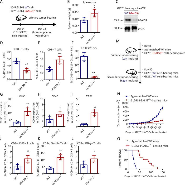
Fig. 6. Effect of exosomal LGALS9 on the mouse CSF immune microenvironment.
a Schematic of the experimental design for (b–i). Briefly, four mice were transplanted with GL-261 LGALS9−/− cells or GL-261 WT cells, followed by immune analysis at 14 days. Spleen size (b) and LGALS9 expression in the CSF (c) of GL-261 LGALS9−/− and GL-261 WT tumor-bearing mice. The percentages of LGALS9hi DCs (d), CD8 + T cells (e), and CD4 + T cells (f) in the CSF of tumor-bearing mice. The expression of MHC I (g), CD40 (h), and TAP1 (i) in DCs in the CSF. The percentage of KI67 + cells among CD8 + T cells (j), the percentage of Gzm B + cells (k) and the percentage of IFN-γ-positive cells (l) in the CSF. m Schematic of the experimental design for n–o. Fourteen mice were transplanted with GL-261 LGALS9−/− cells or GL-261 WT cells, followed by secondary contralateral subcutaneous injection of GL-261 WT cells on day 30. The tumor growth volume (n) and survival rate (o) in the following days. All data are shown as the mean ± SD; *p < 0.05 and **p < 0.01 by a t-test.

