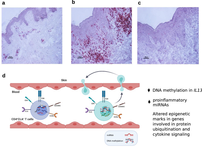
Figure 6.
Identification of CLA+ cells in skin biopsies and epigenetic changes detected in circulating CD4+CLA+ T cells from AD patients that might contribute to skin inflammation. A representative immunohistochemistry staining of the distribution of CLA+ cells in skin biopsies from (a) a healthy control, (b) lesional skin from an AD patient, and (c), rat IgM used as isotype control. Scale bars represent 50 µm. (d) Circulating CD4+CLA+ T cells from AD patients (light blue cell) show significant differences in DNA methylation and miRNA levels compared to CD4+CLA+ T cells from HC (purple cell). The main differences were detected in the reduced DNA methylation of the IL13 gene, the increased expression of proinflammatory miRNAs and coordinated epigenetic changes in genes involved in protein ubiquitination and cytokine signaling in AD patients. Since these CD4+CLA+ T cells can recirculate between skin and blood25, these altered epigenetic marks might contribute to AD immunopathology.

