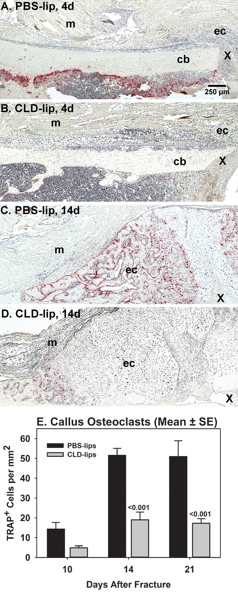
Figure 1. Clodronate liposome treatment reduces fracture callus osteoclasts.

Mouse fracture callus specimens collected at 4, 10, 14, and 21 days after fracture from PBS-lip treated (n= 3, 4, 6, and 6, respectively) and CLD-lip treated (n =3, 5, 6, and 6, respectively) mice were processed for paraffin histology. Shown are sections stained for TRAP activity to identify osteoclasts (red color) at days 4 (A and B) and 14 (C and D) after fracture in PBS-lip (A and C) and CLD-lip (B and D) treated mice. Panels are marked to show the external fracture callus (ec), cortical bone (cb), muscle (m), and fracture site (X). The mean (±SE) number of TRAP+ cells from the 10, 14, and 21 day specimens of the PBS-lip (black) and CLD-lip (gray) treated specimens are shown. Significant differences between treatment groups are noted with associated p values.
