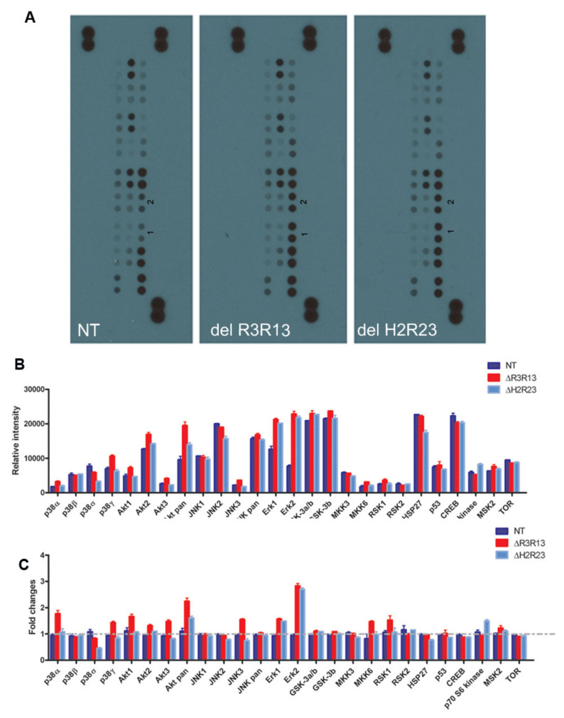
Figure 5.
Signalling pathways involved in the effect of the mini-dystrophins on DMD muscle- derived cells. Non-transduced cells (NT), GFP+ cells purified from LV–SFFV–ΔR3R13–GFP or LV–SFFV–ΔH2R23–GFP-transduced cells were expanded and analysed with a human phosphor-MAPK array kit (A). Data shown are from a 1 min exposure to X-ray film. 1 and 2 in each blot denote the phosph-Erk2 and phosph-Erk1 signals respectively. Relative intensity measurements show expression levels of the 26 MAPKs in each sample (B), as well as by fold changes of each kinase against non-transduced cells in experimental groups (C).

