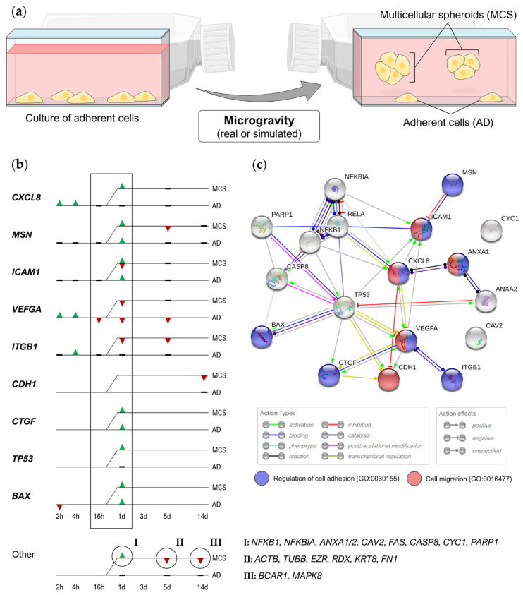
Figure 1.
(a) Under µg conditions, breast cancer cells grow into two distinct populations, characterized by hugely different morphologies. (b) Genes regulated in s-µg-induced multicellular spheroids (MCS) formation of MCF-7 cells [50,51,53]. First MCS were detectable after 24 h of random positioning. ▲, upregulation; ▼, downregulation; (c) STRING (Search Tool for the Retrieval of Interacting Genes/Proteins) interaction network of proteins encoded by the regulated genes. Biological processes that are important both in cancer progression and in MCS formation are colorized. Blue, regulation of cell adhesion (Gene Ontology process GO:0030155); red, cell migration Gene Ontology process GO: 0016477). Gene symbols: ACTB, β-actin; ANXA1/2, annexin 1/2; BAX, Bcl-2-associated X protein; BCAR1, breast cancer anti-estrogen resistance protein 1; CASP8, caspase-8; CAV2, caveolin 2; CDH1, E-cadherin; CTGF, connective tissue growth factor; CXCL8, interleukin-8; CYC1, cytochrome c1; EZR, ezrin; FAS, Fas cell surface death receptor; FN1, fibronectin; ICAM1, intercellular adhesion molecule 1; IKBKG, inhibitor of NF-κB kinase regulatory subunit gamma; ITGB1, integrin-β1; KRT8, cytokeratin; MSN, moesin; NFKB1, nuclear factor kappa B subunit 1; NFKBIA, NF-κB inhibitor A; PARP1, poly [ADP-ribose] polymerase 1; RDX, radixin; TP53, tumor protein p53; TUBB, β-tubulin; VEGFA, vascular endothelial growth factor A. Parts of the figure are drawn using pictures from Servier Medical Art (https://smart.servier.com), licensed under a Creative Commons Attribution 3.0 Unported License (https://creativecommons.org/licenses/by/3.0).

