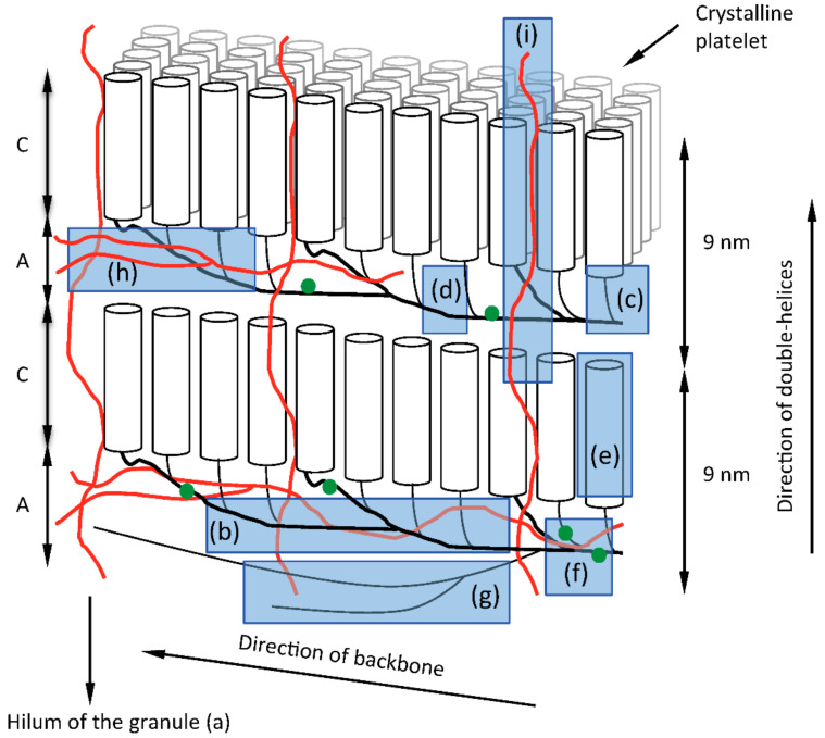
Figure 4.
A cartoon of the building block backbone model of amylopectin with its structural elements in the starch granule along with the possible involvement of amylose. Stacks of amorphous (A) and crystalline lamellae (C) are indicated with a repeat distance of approximately 9 nm. Layers of amylopectin molecules are laid upon each other during the biosynthesis and crystalline platelets are formed by the double-helices (cylinders) as indicated in the 3D-view. The non-reducing ends of the double-helices are directed toward the granule surface and the hilum of the granule (a) is found in the other direction. Amylose (red lines) is suggested to be found interspersed with the backbone of amylopectin (bold black lines) in the amorphous layers, which increases the stability of the starch granules. Some amylose molecules are also crossing the layers, thereby providing additional stability to the layered structure. The structural elements are highlighted in blue as (b) the backbone, (c) building blocks, (d) inter-block segments, (e) external segments (forming double-helices), (f) phosphate groups (green dots), (g) super-long chains, (h) branched amylose, and (i) linear amylose. See also Table 1 which refers to the same structural elements in relation the respective enzymatic reactions responsible for their synthesis. Note that the actual positions of phosphate groups remain uncertain.

