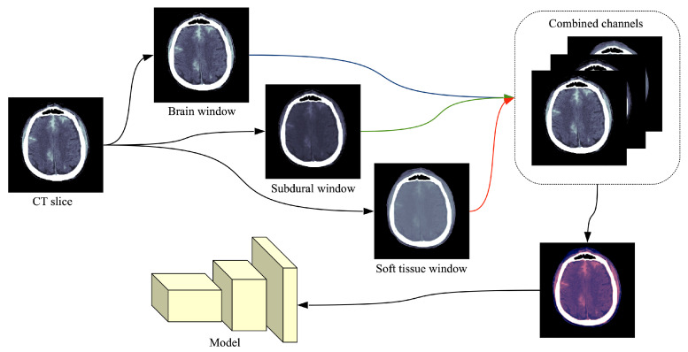
Figure 1.
Preprocessing flow of a single CT slice in DICOM format. Each DICOM file is processed by extracting three different intensity windows (brain window, subdural window, soft tissue window), which are treated as three different channels. The resulting image is what the neural model takes as input. In order to illustrate the final image as an RGB image, we used the following correspondence: brain → red, subdural → blue, soft tissue → green. Best viewed in color.

