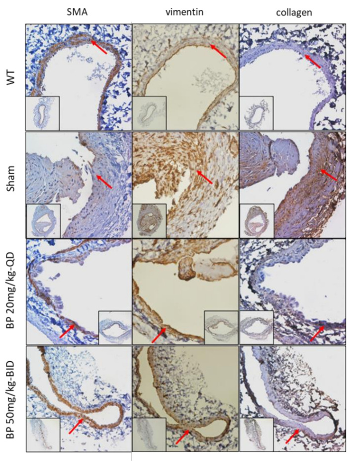
Figure 4.
Immunohistochemical detection (brown color) of a venous limb of AVF from rats treated with different doses of BP for 30 days. The expression of a phenotypic-specific marker (SMA, vimentin and collagen I) in an adjacent section of each group is indicated by a red arrow. Significant inhibition of phenotypic switch was observed in rats treated with BP compared to the control (sham) group. Scale bar = 100 μm.

