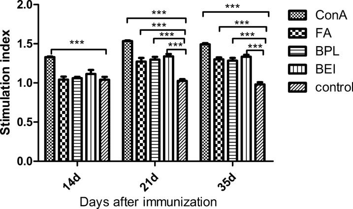
Fig. 4.
The proliferation result of spleen lymphocyte by MTT assay. Spleens of three mice in each group were collected at 14, 21 and 35 dpi, respectively (n = 3). Lymphocytes were obtained and stimulated with inactivated TGEV antigen at 37 °C for 24 h. Con A was used as the positive control, and the DMEM was used as the negative control. Bars represent the mean (± standard deviation) of three replicates per treatment in one experiment. Statistical significance was indicated by ***P < 0.001(extremely significant) compared with the negative control group

