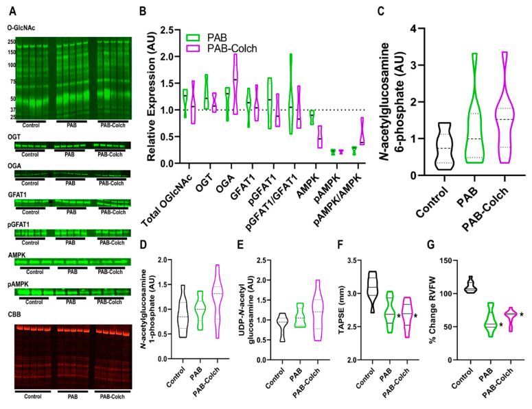
Figure 2.
Colchicine has minimal effects on HBP intermediate levels, O-GlcNAcylation, and right ventricular function in PAB rats. (A) Representative Western blots and subsequent quantification (B) of protein abundance in right ventricular extracts from control, PAB, and PAB-Colch rats (n = 3–5 animals per group). O-GlcNAc (PAB: 1.2 ± 0.2, PAB-Colch 1.1 ± 0.3 fold increase versus control), OGT (PAB: 1.3 ± 0.3, PAB-Colch 1.1 ± 0.2 fold increase versus control), OGA (PAB: 1.2 ± 0.3, PAB-Colch 1.4 ± 0.4 fold increase versus control), GFAT1 (PAB: 1.1 ± 0.3, PAB-Colch 1.1 ± 0.3 fold increase versus control), phosphoGFAT1 (PAB: 1.2 ± 0.4, PAB-Colch 1.0 ± 0.3 relative expression compared to control), pGFAT1/GFAT1 (PAB: 1.1 ± 0.6, PAB-Colch 1.0 ± 0.4 relative expression compared to control), AMPK (PAB: 0.9 ± 0.1, PAB-Colch 0.5 ± 0.2 relative expression compared to control), pAMPK (PAB: 0.2 ± 0.1, PAB-Colch 0.2 ± 0.05 relative expression compared to control), pAMPK/AMPK (PAB: 0.3 ± 0.1, PAB-Colch 0.5 ± 0.3 relative expression compared to control). Quantification of levels of N-acetylglucosamine-6-phosphate (C), N-acetylglucosamine-1-phosphate (D), and UDP-GlcNAc (E) in the right ventricle of control, PAB, and PAB-Colch rats (n = 10 for each group). There were no significant changes in O-GlcNAcylation and HBP intermediates when the three groups were compared. Quantification of right ventricular function shows colchicine did not improve TAPSE (PAB: 2.7 ± 0.2 mm, PAB-Colch: 2.7 ± 0.2 mm) (F) or RV free wall thickening (PAB: 59 ± 16%, PAB-Colch: 65 ± 11%) (G) in PAB rats. (*) indicates significantly different from control as determined by one-way ANOVA with Tukey post-hoc analysis. Tissue was harvested 8 weeks after sham or PAB procedure. CBB: Coomassie brilliant blue.

