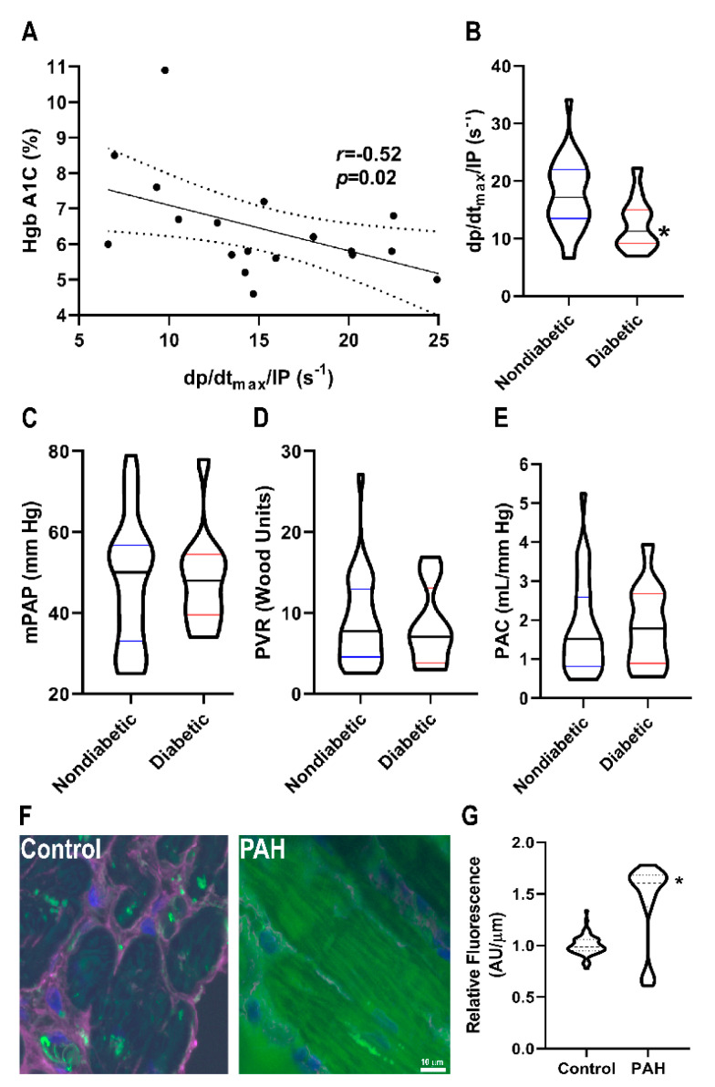
Figure 5.
Relationship between O-GlcNAcylation and RVD in PAH patients. (A) Correlation between HgbA1C and right ventricular contractility in PAH patients. There is a significant negative relationship between HgbA1C and right ventricular contractility (r = −0.52, p = 0.02) (B) Diabetic patients have lower right ventricular contractility than nondiabetic PAH patients (12.5 ± 4.5 s−1 vs. 17.5 ± 6.0 s−1, p = 0.01). There were no significant differences in pulmonary vascular disease severity in diabetic versus nondiabetic patients as quantified by mean pulmonary artery pressure (48 ± 12 vs. 48 ± 16 mmHg, p = 0.37) (C), pulmonary vascular resistance (8.3 ± 4.9 vs. 8.9 ± 5.5 Wood units, p = 0.68) (D), and pulmonary arterial compliance (1.8 ± 1.0 vs. 1.8 ± 1.2 mL/mm Hg, p = 0.64) (E). (F) Representative confocal micrographs of right ventricular sections stained with succinylated wheat germ agglutinin (WGA). (G) Quantification of succinylated WGA signal intensity in right ventricular cardiomyocytes from two control (n = 63 total cells analyzed) and two PAH biopsy specimens (n = 55 total cells analyzed). Green: Succinylated WGA, Blue: DAPI, and Magenta: WGA. * indicates significantly different as determined by t-test in (B) or Mann–Whitney U-test in (G).

