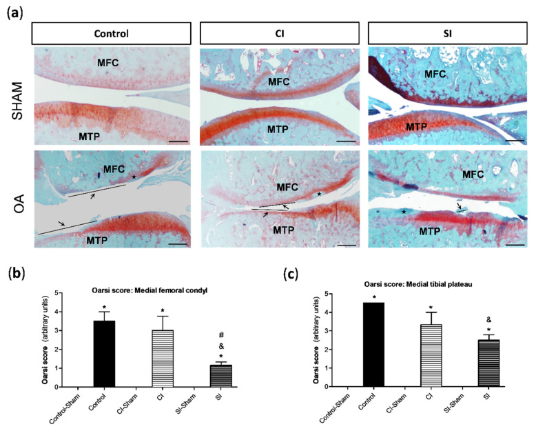
Figure 3.
Histological analysis of articular cartilage. Cartilage lesions were evaluated by the semi-quantitative modified osteoarthritis Research Society International (OARSI) score, as previously indicated in the Materials and Methods section. (a) Representative images of the joint sections stained with Safranin-O-fast green from each group of the study, showing the cartilage of the medial compartment from the tibial plateau (MTP) and femoral condyle (MFC) in the right knee (sham surgery) and left knee (OA surgery). Arrows indicate areas with a loss of cartilage matrix and ★ indicates cartilage with the loss of Safranin staining (indicator of proteoglycan content). Analysis of the semi-quantitative score of the pathological alterations in the cartilage from MFC (b) and MTP (c). Values are mean ± SEM (n = 3 independent animals for each condition). * p ≤ 0.05 vs. the respective sham-operated joint; & p ≤ 0.05 vs. the C group; # p ≤ 0.05 vs. the CI group. Control, non-treated; CI, control injection; SI, H2S donor injection. Scale bar = 500 µm.

