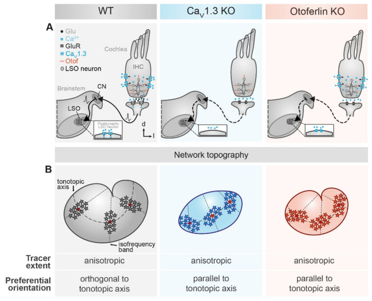
Figure 5.
Summary of LSO astrocyte network topography in mouse models for human hereditary deafness. (A) schematic drawings depicting the subcellular modifications of the different mouse models. Compared to the WT (left), absence of CaV1.3 (middle) and otoferlin (right) from cochlear inner hair cells prevents Ca2+ entry into the inner hair cell and Ca2+ detection, respectively. Subsequently, exocytotic glutamate release is inhibited. Thereby, spontaneous activity of inner hair cells does not result in vesicle fusion, synaptic transmission, and subsequent activation of the auditory pathway. (B) main result of network analysis. LSO astrocyte networks are preferentially anisotropic and oriented orthogonally to the tonotopic axis (left). By contrast, networks in CaV1.3 KO (middle) and otoferlin KO mice (right) are predominantly oriented parallel to the tonotopic axis. Furthermore, the area of the LSO is reduced in both KO models. Moreover, the kidney-like shape of the LSO is lost in the CaV1.3 KO.

