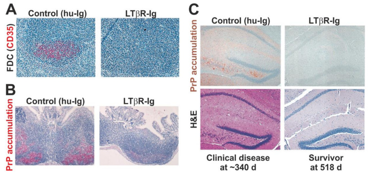
Figure 2.
Oral prion disease pathogenesis is impeded in the transient absence of follicular dendritic cells (FDC) at the time of infection [61]. (A) Immunohistochemical (IHC) analysis shows that the treatment of mice with a soluble lymphotoxin β receptor (LTβR-Ig) transiently ablates FDC (CD35+ cells, red) in secondary lymphoid tissues. (B) Prion accumulation (here shown as disease-specific PrP accumulation by IHC, red) is blocked in Peyer’s patches in the absence of FDC at the time of oral prion infection. (C) Oral prion disease susceptibility is blocked in the absence of FDC at the time of oral prion infection. Upper images show IHC detection of disease-specific PrP (brown) and lower H&E-stained panels show presence of prion disease-specific vacuolation (spongiform pathology) in the brains of clinically-affected control mice. All sections counterstained with haematoxylin (blue). Adapted with permission from the American Society for Microbiology from [61] (J. Virol. 2003; 77:6845–6854. https://doi.org/10.1128/JVI.77.12.6845-6854.2003).

