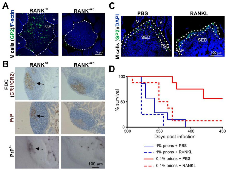
Figure 3.
The density of M cells in the epithelia covering the Peyer’s patches directly influences oral prion disease susceptibility [96]. (A) Immunofluorescence microscopy shows that RANKΔIEC mice specifically lack M cells (GP2+ cells, green) in the follicle-associated epithelia (FAE) of their Peyer’s patches. F-actin, blue. V, villus. (B) In the absence of M cells the accumulation of prions (PrPSc, black and PrP, brown, arrows) upon FDC (CR1/CR2+ cells, brown) in the Peyer’s patches of RANKΔIEC mice is blocked. (C) Immunofluorescence microscopy shows that systemic treatment of C57BL/6J mice with the cytokine RANKL increases the abundance of M cells (GP2+ cells, green) in the FAE. DAPI, cell nuclei, blue. SED, subepithelial dome. (D) RANKL treatment significantly increases susceptibility to oral prion infection by ~10X. PBS/1% vs. RANKL/1%, p = 0.120; PBS/0.1% vs. RANKL/0.1%, p < 0.0078; PBS/1% vs. RANKL/0.1%, p = 0.205; Log-rank [Mantel–Cox] test). Adapted from [96] under the terms of the Creative Commons Attribution Licence (CC-BY-4.0; https://creativecommons.org/licenses/by/4.0/).

