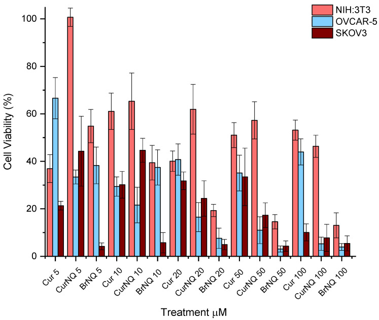
Figure 5.
Cell viability assays were performed using a High Content Imaging System. Three cell lines were utilized, 2 ovarian cancer cell lines namely OVCAR-5 and SKOV3 and 1 healthy fibroblast cell line namely 3T3. Each cell line was treated with varying concentrations of CUR, CurNQ and BrNQ and treatments where incubated for a 24-h interval.

