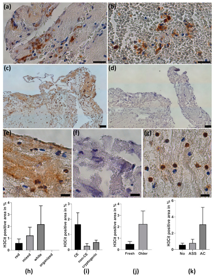
Figure 3.
Neutrophil extracellular traps in thrombemboli. (a,b) Representative H3Cit stained thrombemboli. (a) Magnifications of web-like neutrophil extracellular traps (NETs) within fibrin-rich structures and (b) web-like NETs in RBC-dominating area. (c) Cerebral thrombembolus with a vast amount of H3Cit-positive area and control staining with IgG (d). (e) Magnification illustrates the presence of H3Cit-positive area identified by its brown color in contrast to IgG control (f) and intracellular H3Cit immunoreactivity in neutrophils (g). Counterstaining with hemalum dyes nuclear cells and DNA. H3Cit-positive area was quantified and is presented according to thrombembolus morphology (h), stroke etiology (i), thrombembolus age (j) and pre-existing anticoagulant medication (k). ASA = acetylsalicylic acid, AC = anticoagulation. Scale bars: (a,b) 20 µm, (c,d) 50 µm; (e,f,g) 10 µm.

