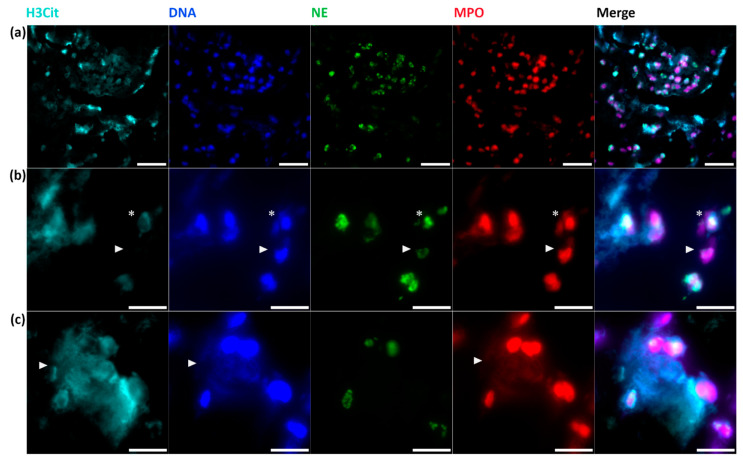
Figure 4.
Immunofluorescent staining of NETs. Immunofluorescent co-staining identifies (a) large thrombembolus area with co-localization of H3Cit, DNA (by DAPI), NE and MPO. Representative magnifications of (b) NETosis (*) and neutrophils without intracellular H3Cit (arrowhead) and (c) web-like NETs with co-localization of extracellular H3Cit, DNA and MPO are shown (arrowhead). Scale bars: (a) 20 µm, (b,c) 10 µm. NE = neutrophil elastase; MPO = myeloperoxidase, DAPI = 4,6-diamidino-2-phenylindole.

