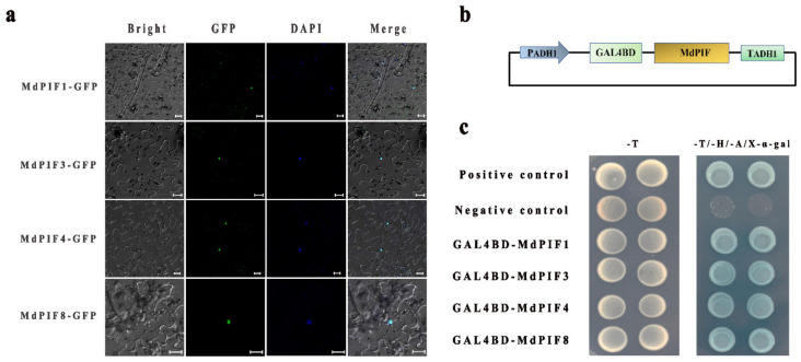
Figure 5.
Subcellular localization and transactivation assay of MdPIF proteins. (a) Subcellular localization of MdPIF proteins. Scale bar = 20 μm. (b) Schematic diagram of the MdPIF-pGBKT7 structure. (c) The construct of pGBKT7-MdPIF was transformed into yeast strain AH109 and examined on SD/-Trp and SD/-Trp/-His/-Ade/X-α-gal plates.

