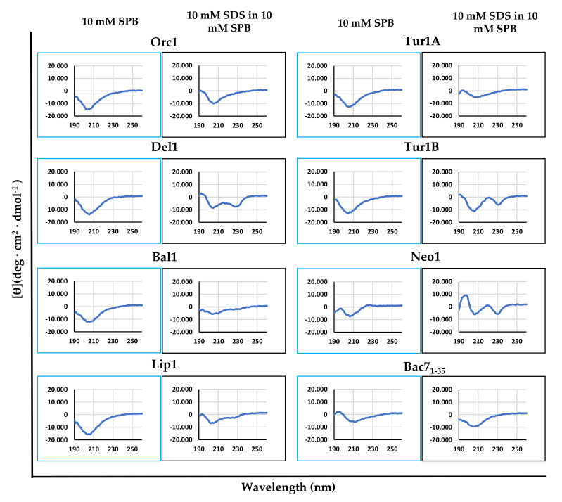
Figure 1.
CD spectra of the peptides (20 µM) in 10 mM sodium phosphate buffer (SPB, first and third columns) and in 10 mM sodium dodecyl sulphate (SDS) in 10 mM SPB (second and fourth columns). The SDS concentration was above the critical concentration for forming micelles, to mimic the anisotropic bacterial membrane environment. Spectra derive from the accumulation of three scans.

