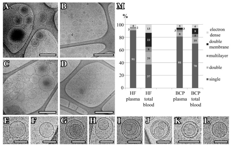
Figure 1.
Representation of exosomes with various morphologies. A–L: Cryo-EM images of exosomes isolated from pooled samples of the plasma of HFs (A), Total blood of HFs (B), plasma of BCPs (C), total blood of BCPs (D), a single vesicle (E,F), double vesicles (G,H), multilayer vesicles (I,J), double-membrane vesicle (I–K), vesicles with an electron-dense cargo in lumen (G,L). Scale bars are 500 nm for micrographs (A–D) and 100 nm for micrographs (E–L). M: Percentages of single, double, double-membrane, multilayer, and vesicles with an electron-dense cargo visualized in the vesicle samples of pooled plasma and total blood from HFs and BCPs.

