Figure 7.
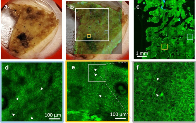
Rapid multi-scale ex vivo MPM imaging with sub-micron resolution to resolve melanocytic dendrites in actinically damaged skin. (a) Photographic image of the excised facial skin tissue. (b) MPM overview map of the skin tissue epidermis generated by strip-mosaic scanning; the 80 MPx image, acquired as a single frame over a 1.2 × 1.0 cm2 area and restored with CARE network in 2.15 min, is illustrated as overlapped with the photographic image of the tissue (opacity 30%). (c) High resolution tile mosaic (6.3 × 6.3 mm2, 49 MPx) acquired in 3 min with 15 accumulated frames for each tile, at 15 μm below the skin surface. (d,e) Digital zoom into the blue and yellow outlined locations in (c), respectively. Images show the distribution of melanocytic dendrites (arrow heads). (f) A close-up of the melanocytic dendrites (white heads) in the outlined area in (e). A z-stack acquired at the same location as the inset in (e) that shows the 3D appearance of the melanocytic dendrites is included in the Supplementary Information (Visualization 3).

