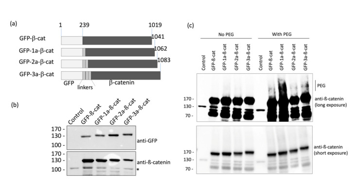
Figure 2.
Design and validation of fluorescent β-catenin fusion proteins. (a) Schematic representation of the different GFP-β-catenin constructs used in this study. (b) HeLa cells were transfected with the various GFP-β-catenin expressing vectors. Cell lysates were analyzed by western blots using anti-GFP and anti-β-catenin antibodies. The asterisk indicates the detection of the endogenous form of β-catenin. (c) Protein extracts of HeLa cells were labeled with 4.4 kDa DBCO-PEG mass tag or incubated with DMSO. β-catenin proteins were detected by Western blot.

