Figure 3.
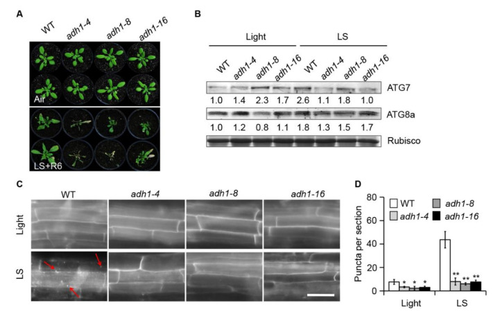
Loss of function of ADH1 attenuates submergence tolerance and autophagosome formation. (A) Phenotypes of 4-week-old wild-type (WT), adh1-4, adh1-8, and adh1-16 plants before treatment (Air) and following 6 d of recovery after 7 d of light (normal light/dark cycle) submergence (LS) treatment (LS+R6). (B) ATG7 and ATG8a protein levels in 4-week-old WT, adh1-4, adh1-8, and adh1-16 plants before treatment (Light) and after 24 h of LS treatment. Coomassie blue-stained total proteins are shown below the blots to indicate the amount of protein loaded per lane. (C) Monodansylcadaverine (MDC) staining of mature root cells from one-week-old WT, adh1-4, adh1-8, and adh1-16 seedlings under normal light/dark (Light) conditions or following 24 h of LS treatment. Red arrows indicate labeled autophagosomes. Bars = 50 µm. (D) Number of puncta per root section in mature root cells from the WT, adh1-4, adh1-8, and adh1-16 seedlings shown in (C). Data are average values ± SD of three biological replicates. For each experiment, 15 sections were analyzed per genotype. Asterisks represent significant differences between WT and adh1 samples, as determined by Student’s t-test (* p < 0.05 and ** p < 0.01).

