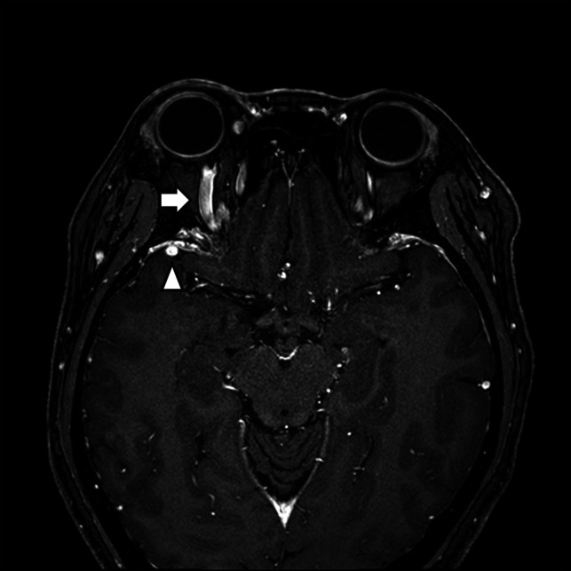
FIG 4.

A CCF case with prominent venous drainage flow in the anterior and lateral venous structures. Axial contrast-enhanced T1-weighted image of a patient with right ocular pain and conjunctival injection. Note the enlarged right superior ophthalmic vein (anterior; arrow) and right sphenoparietal sinus (lateral; arrowhead). The patient was confirmed as having an indirect CCF on digital subtraction angiography.
