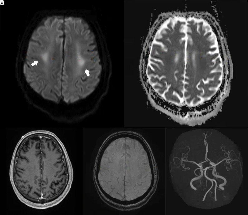
FIG 5.
Brain MR images of a critically ill patient (patient 4) with COVID-19 infection exhibiting impaired arousal and left distal lower extremity paresis. A and B, Paired axial DWI and ADC map show symmetric restricted diffusion of the bilateral perirolandic white matter (arrows). C, Axial contrast-enhanced T1-weighted image at the same level demonstrates no corresponding abnormal enhancement. D, Susceptibility-weighted image at the same level demonstrates no susceptibility effect to suggest hemorrhage. E, 3D reconstruction of the circle of Willis from time-of-flight imaging demonstrates no vascular abnormality.

