
FIG 2.
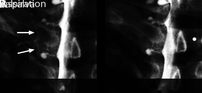
Spot-magnified radiographs of a left T2 nerve root sleeve CSF–venous fistula during an ipsilateral decubitus dynamic myelogram. A, Image acquired during inspiration demonstrates well the contrast-opacified CSF–venous fistula (arrows). B, Image during a Valsalva maneuver results in considerably reduced visualization of the CSF–venous fistula.