
FIG 4.
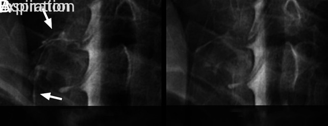
Spot-magnified radiographs of a left C8 CSF–venous fistula during an ipsilateral decubitus dynamic myelogram. A, Image acquired during inspiration demonstrates increased visibility and extent of the CSF–venous fistula (arrows). B, Image acquired during expiration leads to reduced visibility and extent of the CSF–venous fistula.