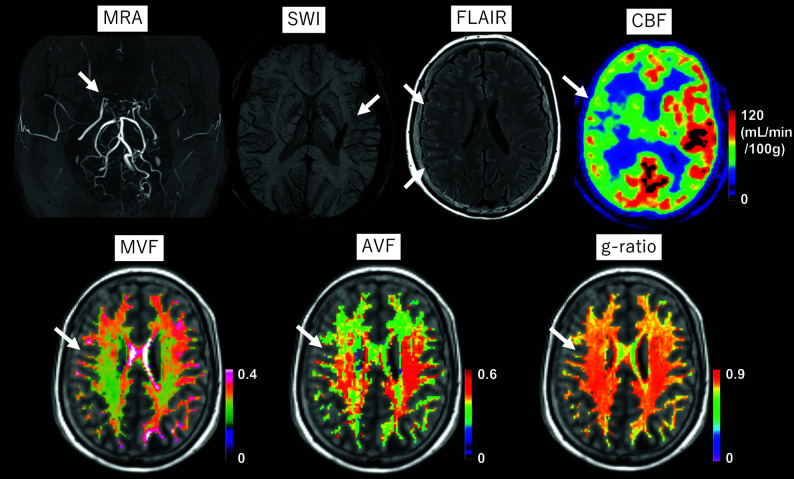
FIG 1.
A 55-year-old female patient with MMD who had a history of thalamic hemorrhage >20 years ago (arrow on SWI). During the annual follow-up, though she remained asymptomatic, the arterial stenosis on the right side gradually progressed (arrow in MRA), and an ivy sign emerged on the right hemisphere (arrows on FLAIR). [15O]-gas PET reveals decreased CBF on the right side (arrow). The myelin volume fraction and axon volume fraction are visually decreased in the right hemisphere (arrows). The right-left difference in the g-ratio (arrow) is not as evident as the differences in the MVF and AVF.

