Figure 1.
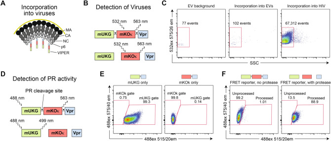
The VIral ProteasE Reporter (VIPER) is incorporated into viral particles and undergoes colorimetric changes in the presence of active HIV protease. (A) Schematic of VIPER reporter incorporation into budding particles. The HIV accessory protein Vpr is packaged into viral particles due to a non-covalent interaction with the Gag p6 structural protein. (B) Schematic of the VIPER reporter construct and detection of viruses. VIPER consists of a mUKG and mKOκ fluorescent pair separated by an HIV protease (PR) cleavage site and fused to the HIV accessory protein Vpr. Viruses can be identified using direct stimulation of the mKOκ subunit using a 532 nm laser, allowing for detection of viral particles by fluorescence thresholding irrespective of PR processing of the reporter. (C) For assessment of specific incorporation of the reporter into viruses, HEK293T cells were transfected with control plasmids (machine noise and EV background), the VIPER construct alone (incorporation into EVs), or the VIPER with HIV (incorporation into HIV). Supernatants were harvested, filtered, and analyzed by nanoscale flow cytometry (NFC). VIPER was robustly and selectively incorporated into HIV particles. (D) Schematic of the detection of HIV PR activity. Stimulation with a 488 nm laser results in differential emission depending on whether the viral PR has cleaved the reporter. (E) Control constructs containing mUKG-Vpr or mKOκ-Vpr alone were generated and used to set gates for nanoscale flow cytometry (NFC) analysis. (F) NFC analysis of viral particles incorporating VIPER demonstrated robust colorimetric changes in the presence of an active HIV protease.

