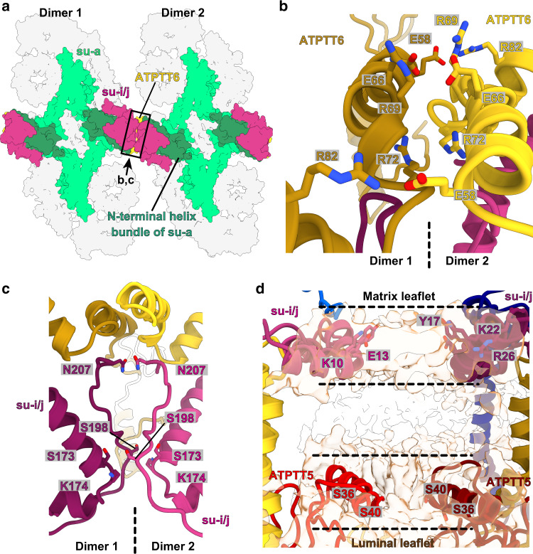
Fig. 7. Type III ATP synthase tetramer interacts via specific protein and lipid-mediated interfaces.
a Luminal view of the tetramer. The N-terminal four-helix bundle of subunit-a (dark green) positions subunit-i/j and ATPTT6 for tetramer interactions. Boxed region and arrow indicate region and view direction shown in close-up view in b, c. b ATPTT6 subunits in the two dimers form electrostatic interactions. c Subunits-i/j in the interface form polar interactions. d Subunits i/j and ATPTT5 interact via bound membrane lipids in the matrix leaflet (black dashed lines) and luminal leaflet (gray dashed lines). Unmodeled lipid density is shown in transparent light brown color.

