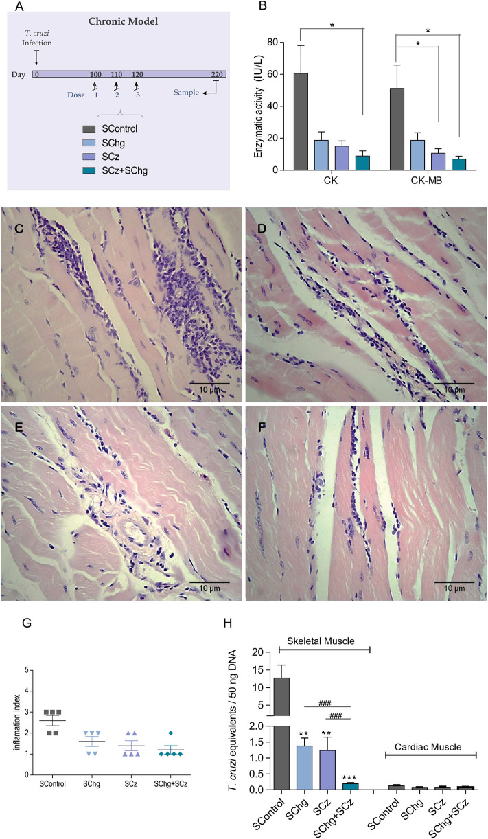
Figure 6.
Prevention of T. cruzi-associated tissue damage by the vaccines administered at the chronic phase of the parasite infection. (A) Scheme of the chronic model of infection: C3H/HeN mice infected with T. cruzi received three doses of the SControl, SChg, SCz, or SChg+SCz on 100, 110, and 120 dpi (n = 5 per group). (B) Enzymatic activity of CK and CK-MB enzymes represented as international units (IU/L). (C,D) Histopathological analysis of skeletal tissue samples at 220 dpi showed (C) confluent foci of mononuclear inflammatory infiltrate with necrosis of the muscle fibers in SControl; (D) isolated foci of mononuclear inflammatory cells with interstitial and perivascular predominance (SChg); (E) nonconfluent mononuclear cells surrounding muscle fibers and interstitial edema (SCz); and (F) isolated mononuclear cells at the interstitial and perivascular level (SChg+SCz). (G) Inflammation semi-quantified and expressed as an index of inflammation: (1) isolated foci; (2) multiple nonconfluent foci; (3) inflammatory confluent foci; and (4) multiple diffuse foci (40). (H) Parasite load in skeletal and cardiac tissues determined by qPCR at 220 dpi. Parasite burden in each tissue was expressed as T. cruzi equivalents per 50 ng of total DNA. Results were referred to a calibration curve previously constructed containing known concentrations of T. cruzi epimastigotes. These results are representative of at least three independent experiments. *p < 0.05, **p < 0.01, ***p < 0.001; ###p < 0.01.

