
Figure 2.
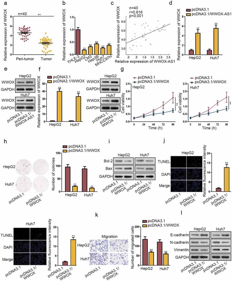
WWOX is positively associated with WWOX-AS1 and exhibits anti-carcinogenic property in HCC. (a) WWOX expression in HCC tissues and adjacent non-tumor tissues was examined through qRT-PCR. (b) WWOX expression in HCC cell lines and one control cell line was detected via qRT-PCR after 48 h of transfection. (c) Pearson’s correlation analysis showed a positive association between WWOX-AS1 expression and WWOX expression in HCC tissues. (d-e) qRT-PCR and western blot were conducted to estimate the effect of WWOX-AS1 overexpression on WWOX mRNA and protein levels after 48 h of transfection. (f) WWOX expression in transfected cells was detected by qRT-PCR and western blot after 48 h of transfection. (g-h) CCK-8 and colony formation assays were utilized to test proliferative ability of pcDNA3.1/WWOX-transfected cells. Cell viability was detected at indicated time points (0, 24, 48, 72 or 96 h). The number of colonies was detected after 14 days of incubation. (i-j) Western blot (after 48 h of transfection) and TUNEL (after 24 h of incubation) were applied to estimated cell apoptosis in transfected cells. (k) Cell migration was measured in WWOX-overexpressed HepG2 and Huh7 cells by transwell assay after 24 h of incubation. (l) The effect of upregulated WWOX on EMT process was tested via western blot analysis of the protein levels of E-cadherin, N-cadherin and Vimentin after 48 h of transfection. **P < .01