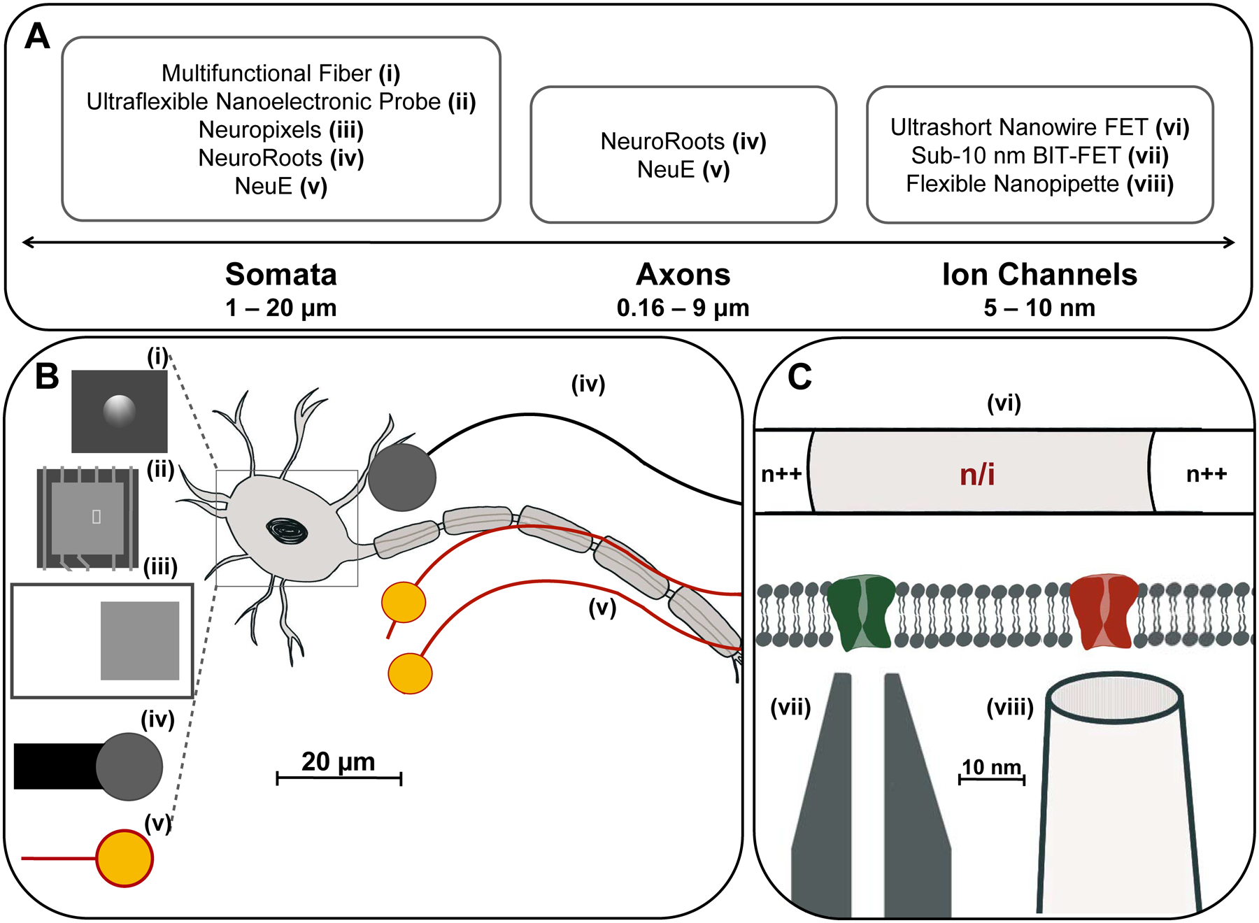
Figure 3.

Bioelectronic neural interfaces inspired by the size and morphology of neuron somata and neurites. (A) Feature sizes of the subcellular structures of the neuron, ranging from a few nanometers for ion channels, 0.16 – 9 microns for axonal diameter, to 1 – 20 microns for neuron somata. (B) Many modern bioelectronic neural interfaces have recording electrodes of similar sizes to neuron somata. Shown are the electrode regions of (i) Multifunctional fibers (d = 5 μm), (ii) Ultraflexible nanoelectronic probe (d = 10 μm), (iii) Neuropixels (d = 12 μm), (iv) NeuroRoots (d = 10 μm), (v) NeuE (d = 8 – 20 μm), where d is the diameter for round electrodes, or width for square electrodes. Additionally, by mimicking the size characteristics of axons, neural interfaces can encourage acceptance in the endogenous neural tissue via promotion of neural progenitor cell migration and integration with the neuronal network. Shown: (iv) NeuroRoots (w ~ 7 μm), (v) NeuE (w = 1 – 4 μm), where w is the width of interconnect ribbons in the electronics. (C) Devices mimicking the size of ion channels, such as the 50 nm ultrashort-channel FET and sub-10 nm BIT-FET, allow recording from individual ion channels with subcellular resolution. Shown: (vi) Ultrashort nanowire FET, (vii) Sub-10 nm BIT FET, and (viii) Flexible nanopipette.
