
Figure 5.
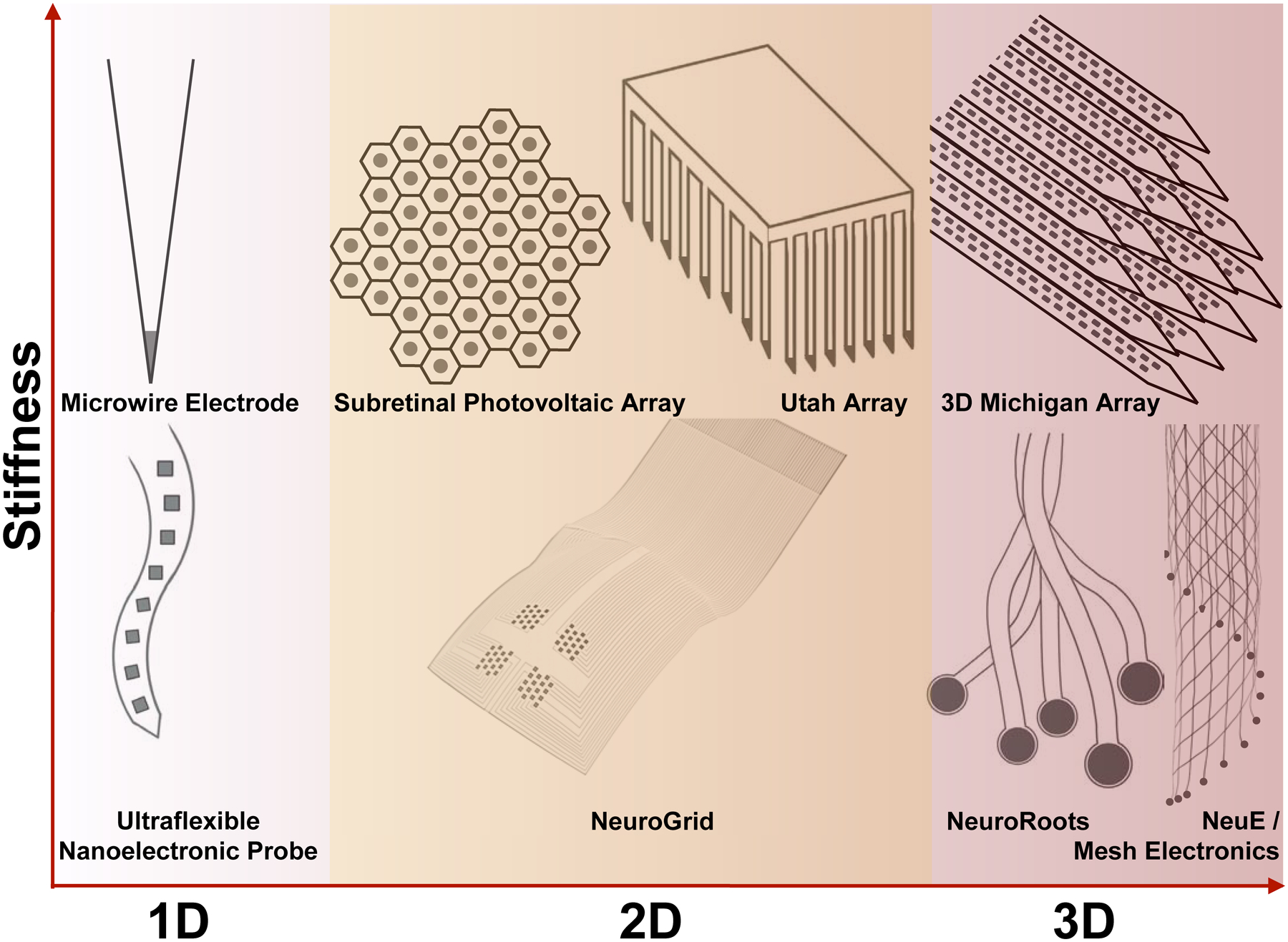
Bioelectronic neural interfaces inspired by the topology of neural tissue. This means that, for neurons within the brain (white-red gradient), neural interfaces constantly advance toward 3D topologies. Starting with the microwire electrode, silicon-based fabrication strategies ignited arraying rigid materials for higher-dimensional interfacing, as seen with the Utah array and the 3D Michigan array. In contrast, interrogation of the neurons in the retina (i.e., retinal ganglion cells) and on the cortical surface requires interfacing at two dimensions on a curvilinear surface (yellow highlight in the middle column). Meanwhile, the joint trend towards higher-dimensional topologies is maintained for flexible bioelectronic neural interfaces, evidenced by the ultraflexible nanoelectronic probe (NET-50), NeuroGrid, NeuroRoots, mesh electronics, and NeuE. Dark gray circles and squares indicate recording sites in these examples. Schematics are not to scale in these drawings.