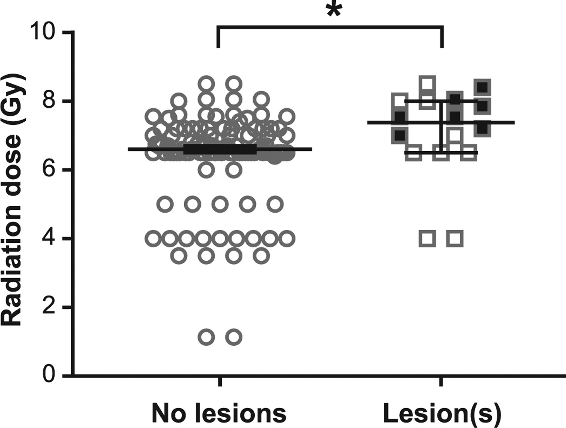
FIG. 2.

TBI is associated with MRI-detectable brain lesions, which occur at higher doses. Animals that developed brain lesions after TBI received higher doses than those without lesions (median: 7.4 Gy vs. 6.6 Gy, respectively; P < 0.02). Filled squares indicate incident lesions which occurred during the surveillance period; these animals received higher doses during TBI (7.8 Gy ± 0.4 SD) than those with lesions present since the time of first evaluation only (6.6 ± 1.5 SD) (P < 0.05). Bars indicate median value, error bars are 95% confidence interval. *P < 0.05.
