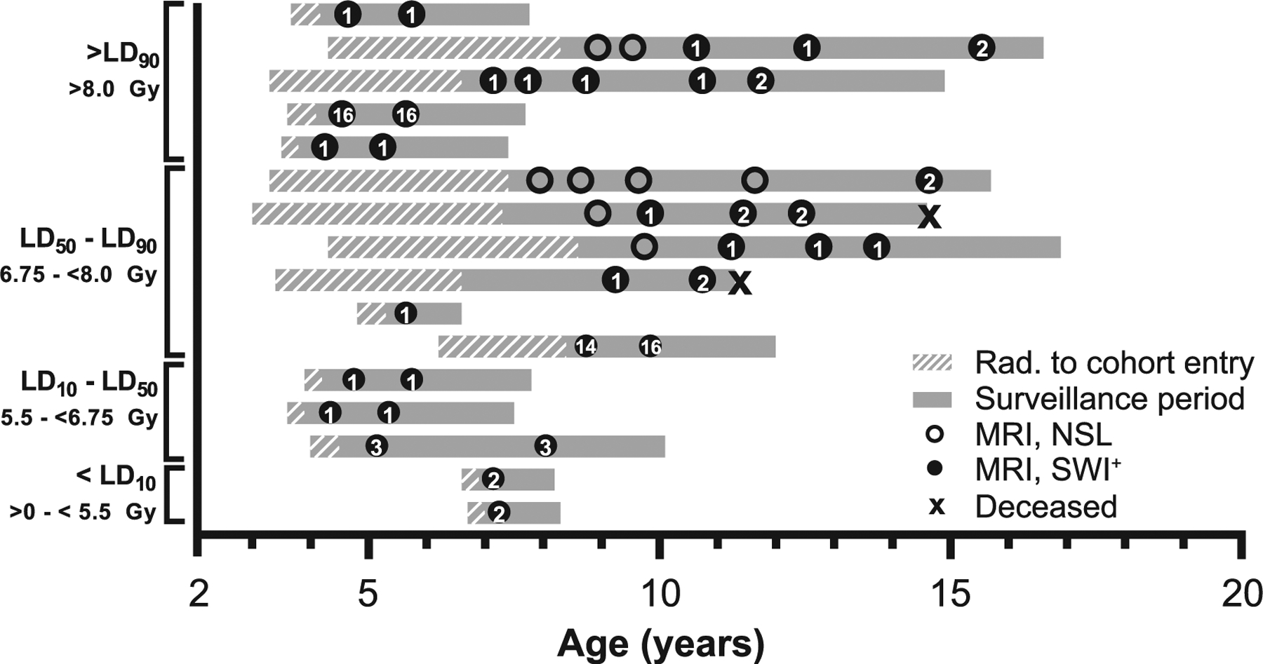
FIG. 3.

Radiologic and life history of animals with SWI-hypointense foci. Each gray bar represents a single animal; animals are listed in order of decreasing radiation dose. Bars begin at age at irradiation; cross-hatched region indicates the period before adoption and cohort enrollment. The solid gray region indicates the period the animal was enrolled within the Radiation Survivors Cohort. Circles indicate brain MRI examinations; open circles represent MRI with no significant lesions (NSL), closed circles represent a SWI lesion that was present. Arabic numerals within closed circles indicate the number of SWI foci at the time of examination. “X” indicates the age at death of deceased animals (n = 2). Lethal dose (LD) stratifications refer to LD30 for the hematopoietic acute radiation syndrome (H-ARS) in rhesus macaques (80).
