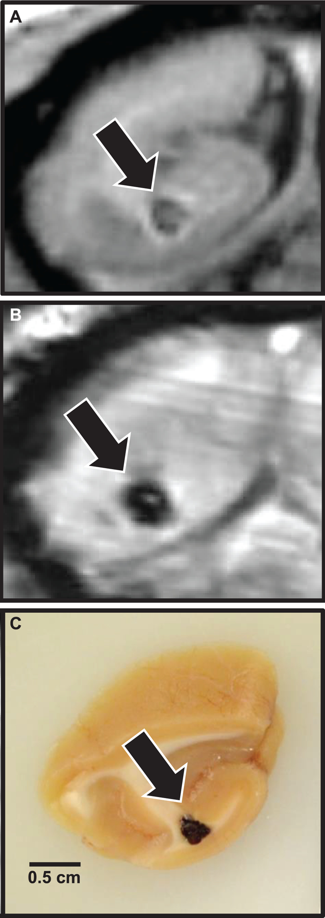
FIG. 5.

Localization of MRI lesions and correlative gross pathology. Coronal sections, occipital lobe, example of a focal brain lesion first noted six years postirradiation, at the time the first MRI scan was completed. Panel A: T1 MRI indicating focal parenchymal loss with central T1 isointense heterogeneity. Panel B: SWI-MRI indicating the presence of iron or calcium (blood or necrosis, respectively). Panel C: Gross tissue, demonstrating focal hemorrhage on sectioned surface.
