
Figure 3. In vivo assessment of cardiac macrophages in newborn mice using 64Cu-Macrin PET.
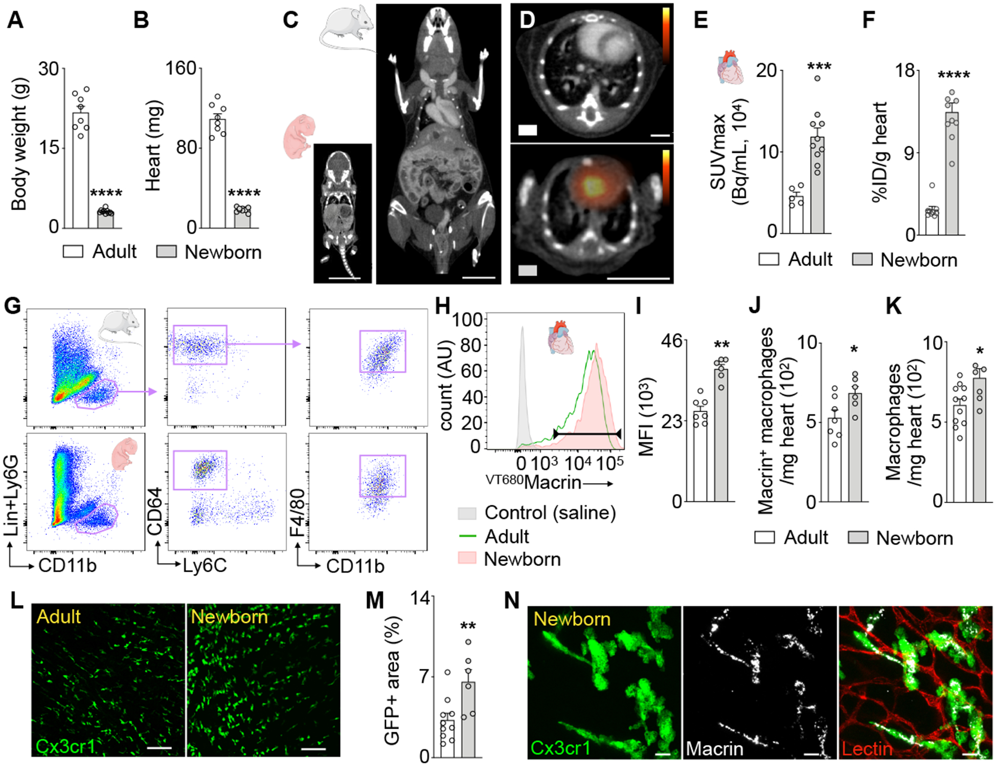
(A) Body and (B) heart weight of adult (9–10 weeks) and 5-day-old mice. (C) CT images of newborn and adult mouse (scale bar, 1cm). (D) PET/CT images visualize 64Cu-Macrin uptake in adult (top) and newborn mouse heart (scale bar, 1cm). (E) 64Cu-Macrin PET quantified as SUVmax and (F) ex vivo scintillation counting. (G) Flow cytometry gating for cardiac macrophages in adult and newborn mice. (H) Histogram showing VT680Macrin uptake in cardiac macrophages. Controls injected with saline (shaded grey), adult (solid green) and newborn mice (shaded orange). (I) Mean fluorescence intensity (MFI) in heart macrophages. (J) Number of VT680Macrin+ macrophages in the heart. (K) Macrophage numbers assessed by flow cytometry. (L) Confocal images of myocardium in Cx3cr1GFP/+ mice (scale bar, 100 μm). (M) GFP+ area in hearts of Cx3cr1GFP/+ mice by confocal microscopy. (N) VT680Macrin (grey) co-localizes with GFP+ macrophages in the heart of a newborn Cx3cr1GFP/+ mouse (scale bar, 10μm). Data are mean ± SEM. *P<0.05, **P<0.01, ***P<0.001, ****P<0.0001.